Making a biological drawing
A biological drawing is a record of what you saw on your own slide. It is not artwork, and it is not a copy of a textbook figure.
Its job is to show the shapes, proportions and contents of the cells in front of you, accurately enough that someone else could recognise the tissue from it.
The conventions
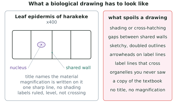
- Pencil only, sharp. Pencil can be corrected and gives one clean line.
- One continuous line for each edge. Sketchy, hairy or doubled outlines make it impossible to tell where a wall actually is.
- No shading, no stippling, no cross-hatching, no colouring in. Depth and tone are not information here.
- Draw it large — at least half the space you have given it. Detail you can see at the microscope disappears in a small drawing.
- Title it with what the material is and how it was viewed, for example "Lower epidermis of harakeke leaf".
- State the magnification you viewed it at, as ×400.
- Label lines ruled, drawn with a ruler, level, and never crossing each other. Each line touches the structure it names. No arrowheads.
- Write labels outside the drawing, lined up down one side, not squeezed inside a cell.
Getting the drawing accurate
Following the conventions makes a drawing tidy. Getting it accurate is a separate job.
- Recognisable shape. A drawn cell should be the shape the real one was — brick-like, column-like, curved, or irregular. If a reader cannot tell which tissue it came from, the shape is wrong.
- Correct proportions. If the real cells are three times longer than they are wide, the drawn ones must be too. A row of squares standing in for long rectangles is a proportion error, not a tidiness one.
- No gaps between neighbouring cells. Cells in a plant tissue share their walls. White space between them says there is a gap, and there is not.
- The organelles you could see, at the magnification you used. A stained nucleus, chloroplasts and a large vacuole belong on the drawing if you saw them. Ribosomes never do, at any magnification.
- Draw a few cells well, not the whole field badly. Two or three cells drawn accurately show more than twenty drawn as a grid.
Annotating the drawing
A label names a structure. An annotation is a short note beside the label saying something about it.
- Annotate a specialised feature with what it does — "guard cell, wall thicker on the inner side".
- Keep annotations short and keep them outside the drawing.
- Annotate what you can see in your drawing. A note about a structure you did not observe belongs nowhere on it.
Three specimens, three drawings
- One drawing for each specimen — the two plant tissues and the unicellular organism.
- Made over separate sessions, so each drawing is done at the microscope with that slide in front of you, not reconstructed later from memory.
- Date each one and keep them together. A drawing you cannot match to a slide is a drawing of nothing in particular.